
CT Scanner 16 -Slice HD Spiral Medsinglong Mslctx04
MSLCTX04Medsinglong 16-Slice HD Spiral CT ScannerImages shot by it Parameter : No.Technical featureDescription1Gantry1.01Gantry typeLow voltage slip-ring withAccuSlip-ring technology1.02Gantry driven typeStrap-driven1.03Patient opening70cm1.04Gantry tilt modeDual-mode gantry tilt1.05Mechanical til......
Send Inquiry
Product Description
MSLCTX04
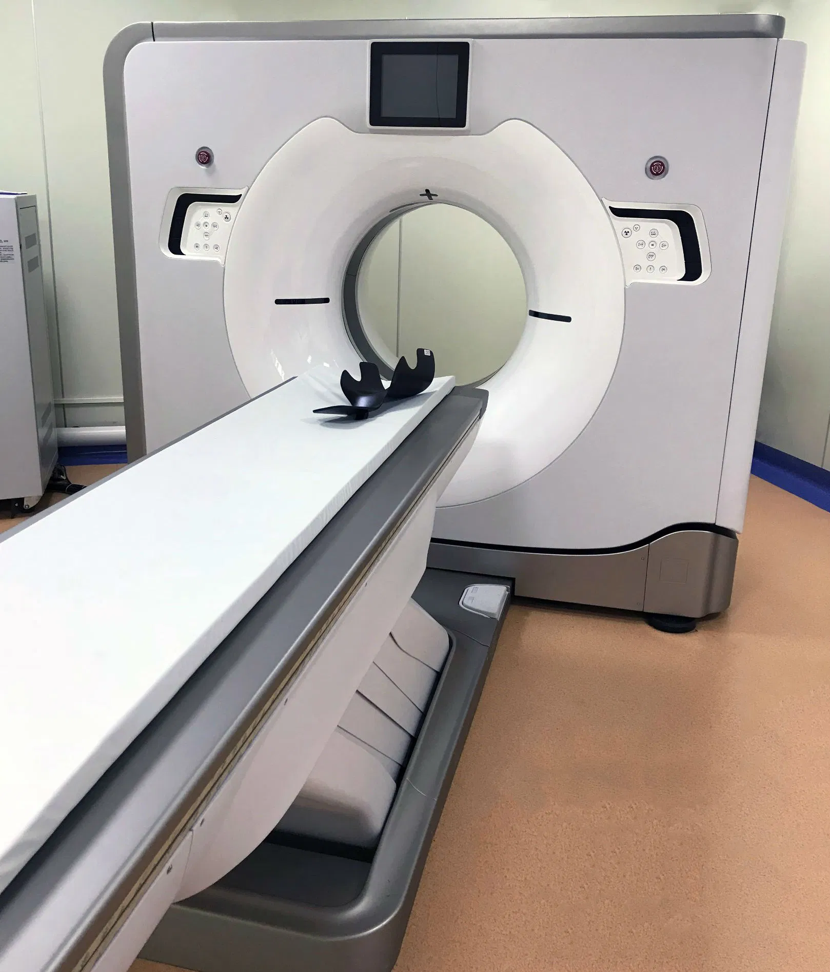
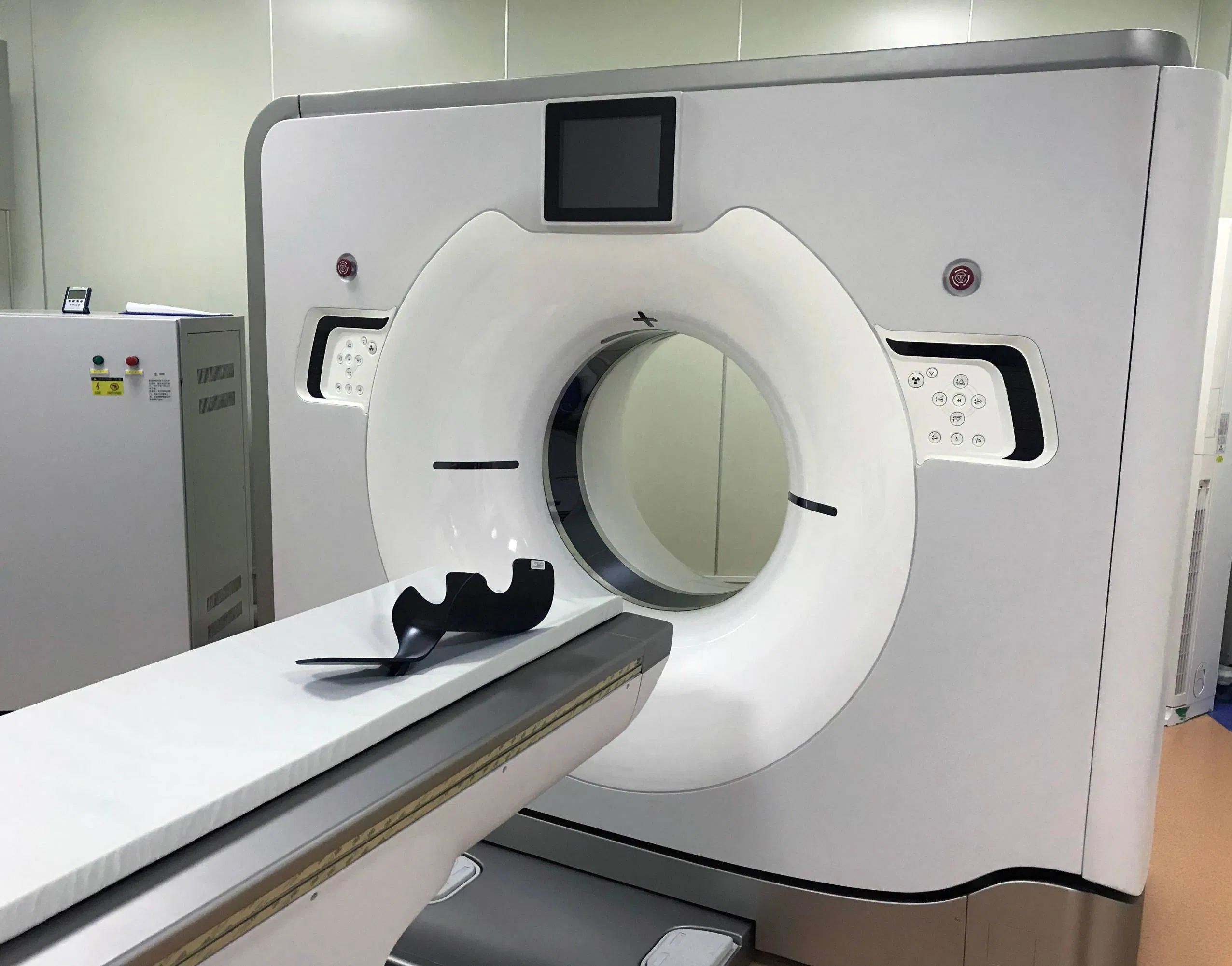
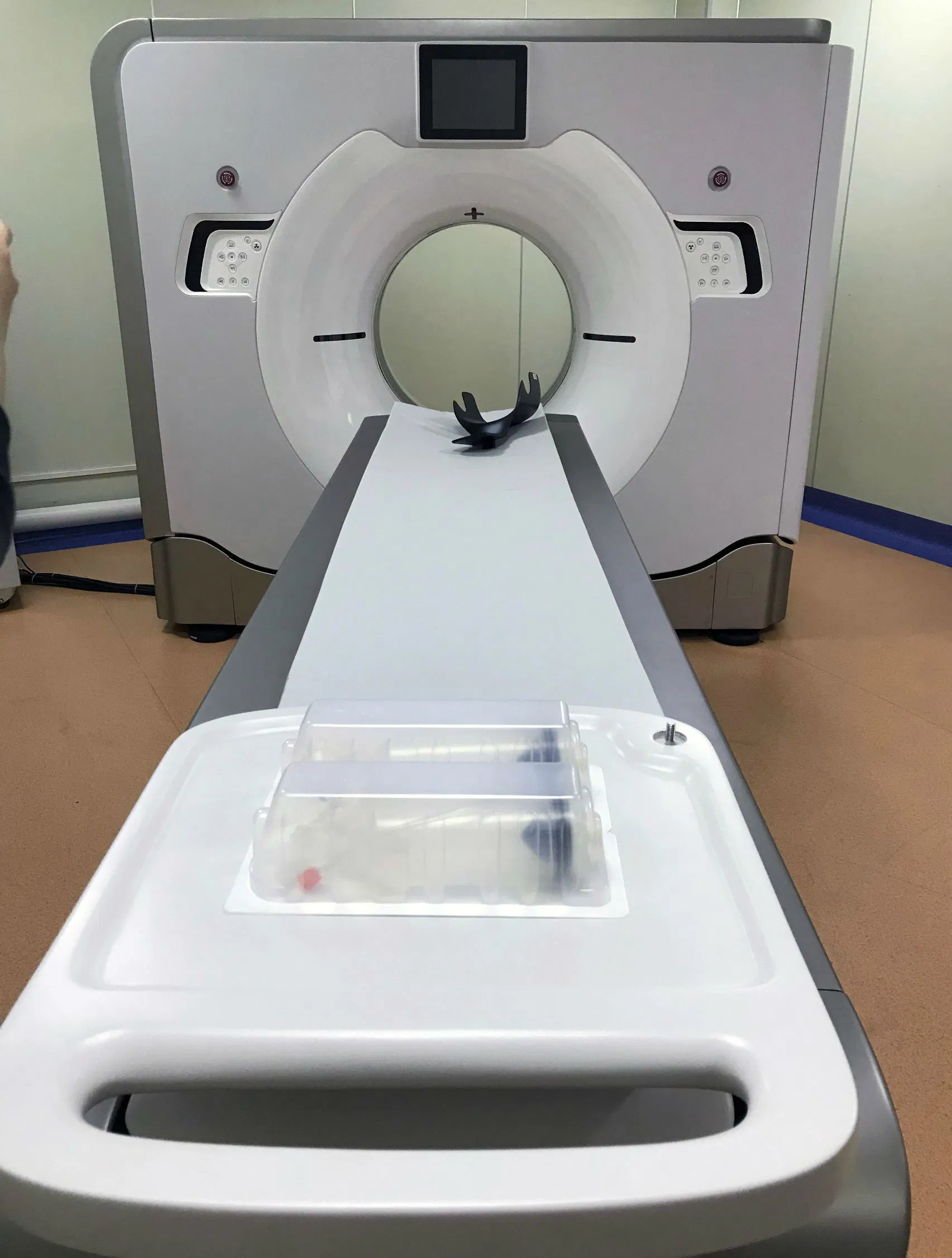
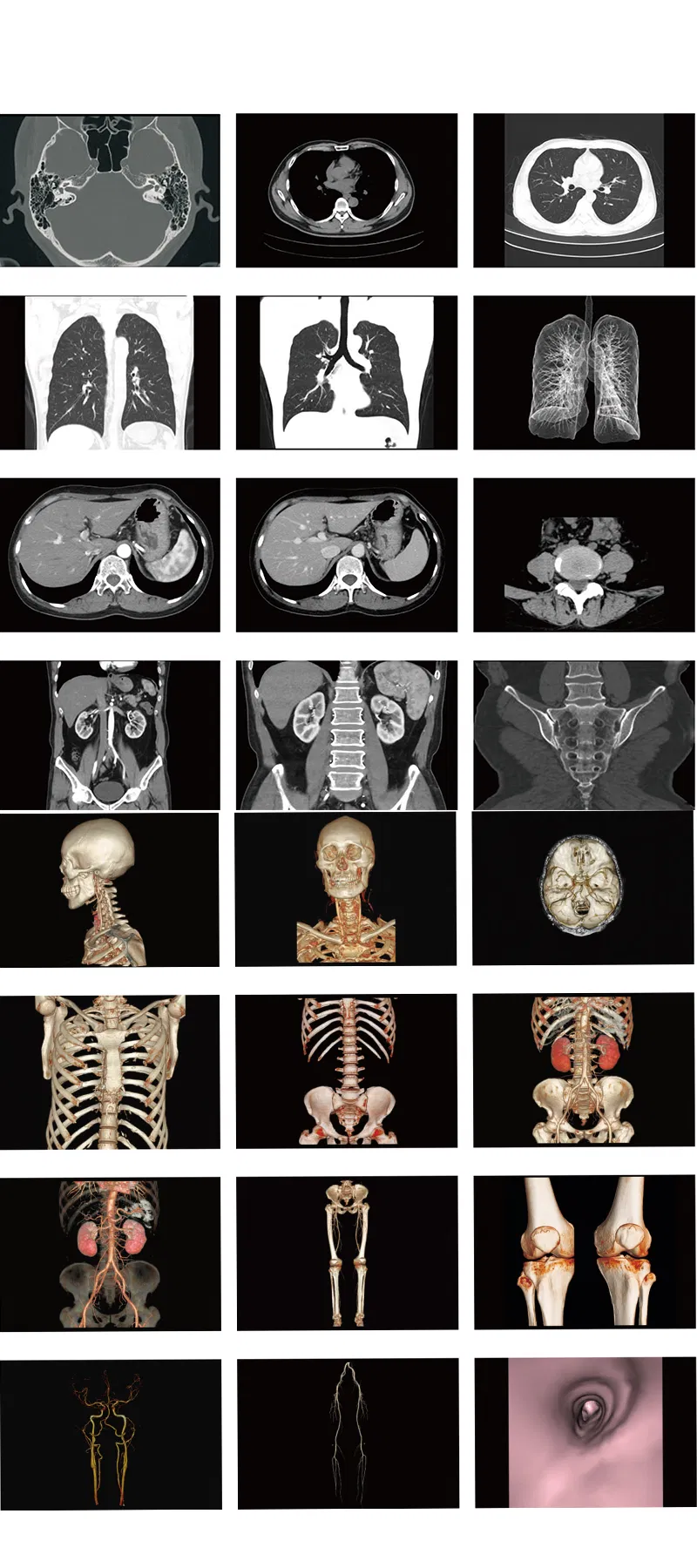
Medsinglong 16-Slice HD Spiral CT Scanner
Images shot by it
Parameter : Medsinglong 16-Slice HD Spiral CT Scanner
Images shot by it
| No. | Technical feature | Description |
| 1 | Gantry | |
1.01 | Gantry type | Low voltage slip-ring with AccuSlip-ring technology |
| 1.02 | Gantry driven type | Strap-driven |
| 1.03 | Patient opening | 70cm |
| 1.04 | Gantry tilt mode | Dual-mode gantry tilt |
| 1.05 | Mechanical tilt capability | ±30° |
| 1.06 | Digital tilt capability | ±50° |
| 1.07 | Gantry remote-Control | Provided |
| 1.08 | Detector type | OptiWave rare-earth ceramic detector |
| 1.09 | Numbers of detector rows | 32 |
| 1.10 | Width of Z-axle detector | 20mm |
| 1.11 | Detector columns of channels per row | 912 |
| 1.12 | Numbers of detector columns | 29184 |
| 1.13 | Data-transfer type | RF,optical fiber communication |
| 1.14 | 3D laser orientation | Provided |
| 1.15 | External X-ray enable | Interface for Foot-Pedal Provided |
| 1.16 | Automatic exposure control(mA Modulation) | Provided |
1.17 | Auto-voice manager | Breath Graphical Display Hold Message (Record/Playback) Breath Message(Record/Playback) |
| 1.18 | Eenergy conservation management | Provided |
| 1.19 | Acquisition mode | 16 × 0.625mm, 16 × 1.25mm |
2 | Scan parameter | |
| 2.01 | Shortest 360 degree rotation time | 0.5s |
| 2.02 | Allowed rotation times | 0.5s,0.8s,1.0s,1.5s,2.0s |
| 2.03 | Slice numbers per rotation | 16 |
| 2.04 | Minimum slice thickness of scan | 0.625mm |
| 2.05 | Minimum slice thickness of reconstruction | 0.625mm |
| 2.06 | Maximum slice thickness of scan | 10mm |
2.07 | Nominal reconstruction slice thickness | 0.625mm,1.25mm,2.5mm,5.0mm, 7.5mm,10mm |
| 2.08 | Speed of image reconstruction(512×512) | 65 frames/s |
| 2.09 | Scan FOV | 52cm |
| 2.10 | Image reconstruction matrix | 512×512,1024×1024 |
| 2.10 | Image reconstruction matrix | 512×512,1024×1024 |
| 2.11 | Image display matrix | 512×512,1024×1024 |
| 2.12 | Maximum continuous scan duration | 120s |
| 2.13 | Maximum continuous scan length | 180cm |
| 2.14 | Direction of TOPO | Front-back,Left-right |
| 2.15 | Max. length of TOPO | 180cm |
| 2.16 | Range of pitch | 0.5~1.5 |
2.17 | Scan mode | Scout scan Axial scan Helical scan Cine scan |
3 | HVPS and Tube | |
| 3.01 | Maximum continuous output of HV generator | 50kW |
| 3.02 | Tube kV selections | 80kV,100 kV,120 kV,140 kV |
| 3.03 | Tube mA range | 10~420mA |
| 3.04 | Tube anode heat capacity | 5.0MHU |
| 3.05 | Heat dissipation rate | 815kHU/min |
| 3.06 | Type of cooling | Oil cooling + Air cooling |
3.07 | Tube focus | Large:1.0 mm×1.0mm Small:0.5mm×1.0mm |
| 3.08 | Dynamic flying focal spot technology | Provided |
| 4 | Patient table | |
| 4.01 | Maximum horizontal-movable range | 1850mm |
| 4.02 | Table horizontal-scannable range | 1800mm |
| 4.03 | Table horizontal-position repeatability | ±0.25mm |
| 4.05 | Maximum vertical-movable range | 500mm |
| 4.06 | Maximum speed of vertical movement | 20mm/s |
| 4.07 | Maximum speed of horizontal movement | 150mm/s |
| 4.08 | Maximum patient weight | 205kg |
| 4.09 | Foot pedal of patient table control | Provided |
5 | Image Quality | |
| 5.01 | High contrast resolution | 21lp/cm@0%MTF |
| 5.02 | Low contrast resolution | [email protected]% |
| 5.03 | Isotropic imaging resolution | 0.625mm |
| 5.04 | Range of CT numbers | -32767~32768 |
| 5.05 | Image noise | ≤0.25@28mGy |
| 6 | Computer subsystem | |
| 6.01 | CPU | 3.5GHz |
| 6.02 | Memory | 16GB×4 |
| 6.03 | Storage of hard-disk | 1T×2 |
| 6.04 | Monitor | 24'' LCD Monitor |
| 6.05 | Resolution of monitor | 1920×1200 |
| 6.06 | Image-data external storage type | CD/DVD/USB |
| 6.07 | Time of image reconstruction(512×512) | 15.4ms/frame |
| 6.08 | DICOM 3.0 interface | Provided |
| 6.09 | Printer DICOM 3.0 interface | Provided |
| 6.10 | Auto filming | Provided |
| 6.11 | Worklist function | Provided |
7 | Advanced application | |
| 7.01 | Multi-Planar Reconstruction(MPR) | Provided |
| 7.02 | Curve Multi-Planar Reconstruction(CPR) | Provided |
| 7.03 | Surface Shaded Display(SSD) | Provided |
| 7.04 | Volume Rendering(VR) | Provided |
| 7.05 | Maximum Intensity Projection(MIP) | Provided |
| 7.06 | Minimum Intensity Projection(MinIP) | Provided |
| 7.07 | Virtual Endoscopy(VE) | Provided |
| 7.08 | CT angiography(CTA) | Provided |
| 7.09 | Tissue segmentation | Provided |
| 7.10 | One click bone remove | Provided |
| 7.11 | One click patient table remove | Provided |
| 7.12 | Bolus-tracking Technology | Provided |
| 7.13 | Spiral auto start | Provided |
| 7.14 | Cine display | Provided |
| 7.15 | AbastTM bone artifact suppression technology | Provided |
| 7.16 | AmastTM metal artifact suppression technology | Provided |
7.17 | Admir3D fulll-domain iterative reconstruction | Provided |
| 7.18 | Low-dose pediatric scan technology | Provided |
| 7.19 | Low-dose lung scan technology | Provided |
7.20 | AccuHead grey-white matter enhanced technology | Provided |
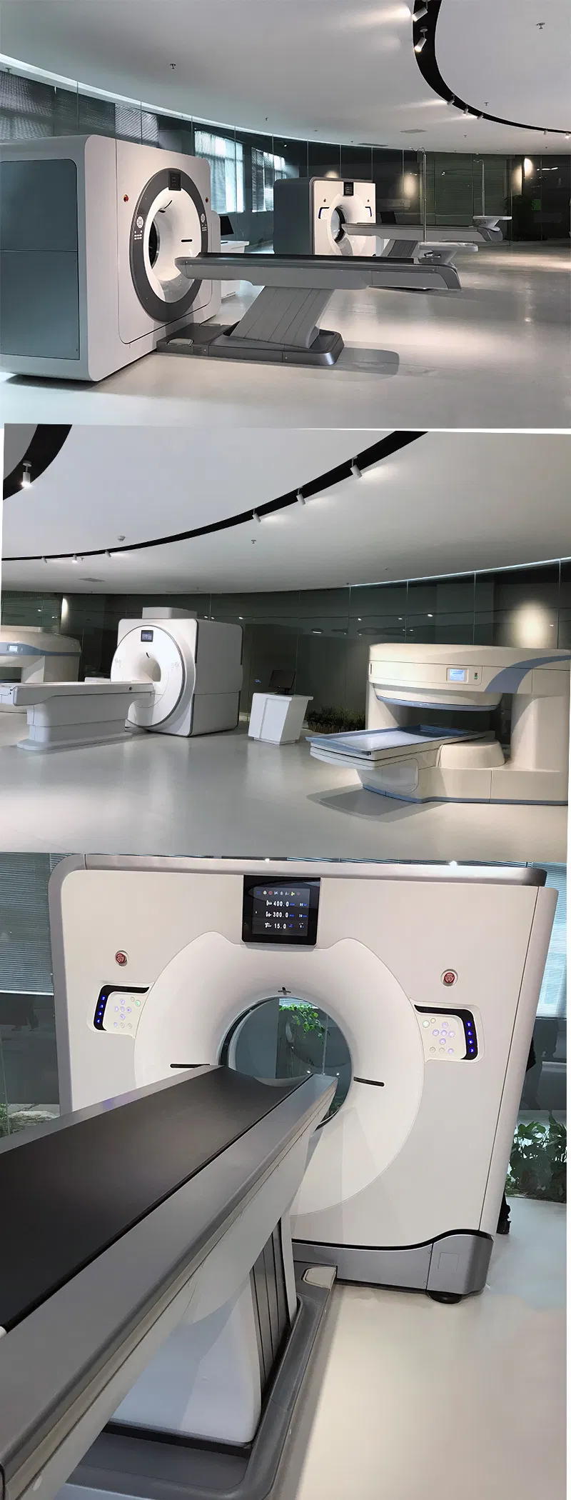
We sincerely invite you to visit our workshop
and experience functions of the machine in person.
(Please confirm the schedule with LeBron , by Email )
Let's meet each other.
Related Category
Beauty Equipment
Medical Diagnosis Equipment
Dental Equipment & Supplies
Physical Therapy Equipment
Disinfect Equipment & Sterilizer
Send Inquiry
Please Feel free to give your inquiry in the form below. We will reply you in 24 hours.


